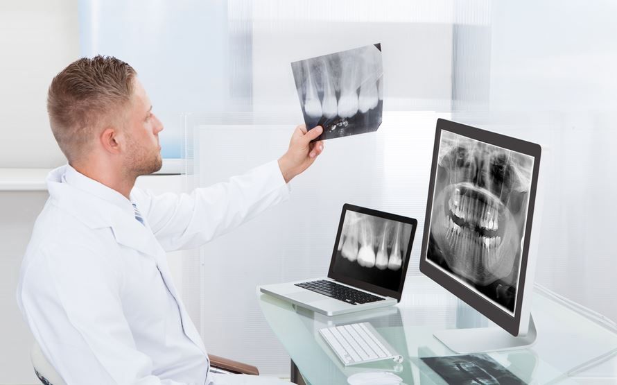
A panoramic dental X-ray provides a comprehensive view of the entire mouth, capturing the teeth, jaws, and surrounding structures in a single image. This type of imaging is particularly valuable for orthodontic evaluations because it allows for an overall assessment of alignment, growth patterns, and potential issues that might affect treatment planning. Unlike traditional Panoramic Dental X-Ray Cost in Dubai that focus on small sections, panoramic imaging offers a broad perspective, making it easier to identify concerns that could impact orthodontic outcomes.
Role in Orthodontic Assessments
Panoramic X-rays play a crucial role in orthodontic evaluations by giving a clear overview of the patient’s dental structure. They allow practitioners to analyze the positioning of teeth and detect issues that might not be visible during a standard oral examination. This imaging helps in identifying crowded teeth, missing teeth, impacted teeth, and other irregularities. By providing a detailed map of the dental and skeletal structures, panoramic X-rays support more accurate treatment planning and monitoring of progress over time.
Identifying Dental and Skeletal Issues
Panoramic dental X-rays are essential for spotting underlying dental and skeletal issues that can influence orthodontic treatment. They can reveal the presence of extra teeth, tooth impactions, and the overall development of jaw bones. They also assist in evaluating the relationship between the upper and lower jaws, which is critical when planning corrective procedures. Through this comprehensive imaging, potential complications can be anticipated early, reducing the likelihood of surprises during treatment.
Advantages Over Traditional X-Rays
Panoramic X-rays offer several advantages over traditional dental X-rays when it comes to orthodontic evaluations. The wide coverage allows for a single image that captures all teeth and jaw structures, eliminating the need for multiple individual X-rays. This approach is less time-consuming and more efficient. Additionally, the detailed visualization aids in diagnosing structural abnormalities and planning interventions with greater precision. The non-invasive nature of the procedure also contributes to a more comfortable experience for patients undergoing orthodontic assessments.
Factors Affecting the Imaging Process
The quality and effectiveness of a panoramic X-ray for orthodontic purposes can be influenced by several factors. Proper positioning of the patient is essential to ensure that the image accurately reflects dental and skeletal structures. Equipment calibration, image resolution, and the experience of the technician can also impact the clarity and usefulness of the results. Ensuring optimal conditions during imaging contributes to a more reliable evaluation and supports effective treatment planning.
Frequency of Panoramic X-Rays in Orthodontics
During orthodontic treatment, panoramic X-rays are often taken at key intervals to monitor progress and adjust treatment plans. The initial X-ray provides a baseline for assessing dental alignment and jaw development. Subsequent images may be used to track tooth movement, monitor growth patterns, and confirm that treatment is progressing as planned. Regular imaging allows for timely interventions if adjustments are needed, enhancing the efficiency and outcomes of orthodontic care.
Enhancing Treatment Planning
Panoramic X-rays significantly enhance treatment planning by providing a comprehensive understanding of the patient’s oral anatomy. The images allow orthodontic professionals to determine the best course of action, whether it involves braces, aligners, or other corrective measures. By visualizing the full dental arch, the position of each tooth, and jaw alignment, treatment plans can be customized to meet the individual needs of each patient. This level of detail supports more predictable and successful orthodontic outcomes.
Benefits for Early Diagnosis
Early diagnosis of dental or skeletal issues is another major benefit of panoramic X-rays. Detecting problems before they progress allows for interventions that can prevent more complex treatment later. For young patients, this can mean guiding jaw growth and alignment effectively, potentially reducing the need for extensive procedures in the future. Early evaluation also helps identify hidden issues such as impacted teeth or developmental anomalies that may otherwise go unnoticed.
Addressing Complex Cases
For complex orthodontic cases, panoramic X-rays provide indispensable information. They allow practitioners to assess not only the teeth but also the surrounding bone structure and sinus areas. This is particularly important when planning treatments that involve significant movement of teeth or adjustments to the jaw. By having a complete view, potential complications can be anticipated, and treatment strategies can be refined to achieve the desired results efficiently and safely.
Common Questions
Why are panoramic X-rays preferred for orthodontics?
They provide a complete overview of the teeth and jaw, allowing for accurate diagnosis and treatment planning.
How often should these X-rays be taken?
They are typically used at the start of treatment and periodically throughout the process to monitor progress and make necessary adjustments.
Are panoramic X-rays safe for repeated use?
Yes, modern imaging uses minimal radiation, and the procedure is considered safe when conducted appropriately.
Can panoramic X-rays detect hidden dental issues?
Yes, they can reveal impacted teeth, misalignments, and other structural abnormalities not visible during a routine exam.
How do these X-rays improve treatment outcomes?
By providing detailed images, they enable customized and precise treatment plans, reducing the likelihood of complications and enhancing efficiency.
Panoramic Dental X-Ray Cost are an invaluable tool in orthodontic evaluations. Their ability to provide a comprehensive view of dental and skeletal structures supports accurate diagnosis, early intervention, and effective treatment planning. The clarity and breadth of these images make them a cornerstone of modern orthodontic care, helping ensure predictable and successful outcomes for patients undergoing treatment.